Detailansicht
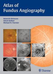
Atlas of Angiography of the Optic Fundus
ISBN/EAN: 9783131405517
Umbreit-Nr.: 1609090
Sprache:
Englisch
Umfang: 192 S., 638 Illustr.
Format in cm:
Einband:
gebundenes Buch
Erschienen am 19.04.2006
Auflage: 1/2006
- Zusatztext
- Angiography of the ocular fundus is a standard examination method that should be mastered by every ophthalmologist treating posterior segment diseases.Outstanding pictures - concise text - Description of the most relevant disease entities seen in daily practice - Double-page layout - Excellent angiographic photo documentation - Combined with significant comments on pathogenesis, indications for angiography, additional diagnostic examinations and decision making Your advantages: - The latest classifications of early and late AMD - Learn standard angiographic methods - Search for the most important angiographic patterns - Interpret angiographies confidently - Follow-up on recent AMD treatment regimens including intravitreal injections of VEGF-antagonists Uptodate application and further developments of standard techniques: Fluorescein angiography Indocyanine angiography Stereoangiography Use and limitations of evolving techniques: - Fundus autofluorescence - Infrared reflectance imaging - Wide-angle imaging Benefit from the experience of renowned lecturers in varying specialities!
- Kurztext
- An essential reference for the classroom and the clinic In modern ophthalmology, angiography of the ocular fundus is one of themost important techniques available for the diagnosis of posterior segment diseases. It is critical that every clinician knows how to recognize significant patterns and confidently interpret the results. The Atlas of Fundus Angiography is the most complete compilation of information on this technique, presented here in beautifully illustrated detail. Key features: Large format and 638 outstanding illustrations with concise commentary for each condition. A stateoftheart overview and comparison of methods, including fluorescein angiography, indocyanine green angiography, fundus autofluorescence, stereoscopic angiography and optic coherence tomography. Logical, didactic structure and accessible layout that teaches you how to seek out important angiographic patterns and reach a sound diagnosis. Systematic coverage of epidemiology, pathophysiology, clinical presentation, diagnostic approach and therapy of the most common chorioretinal disorders including agerelated macular degeneration, diabetic macular edema, and retinal venous occlusion, as well as other vascular diseases, hereditary and toxic retinal diseases, tumors, inflammatory and autoimmune diseases, and disorders of the optic nerve head. Pre and posttreatment retinal imaging in current and evolving therapeutic methods including photodynamic therapy and intravitreal injection of corticosteroids or antiangiogenetic drugs. Insightful assessments of evolving techniques and their uses and limitations, including fundus autofluorescence, infrared reflectance imaging, and wideangle imaging. Expert contributors from all relevant subspecialties. Uptodate and lavishly illustrated, the Atlas of Fundus Angiography is an indispensable tool for clinicians of all levels seeking to master this fundamental technique.
- Autorenportrait
- Heimann, Kellner, Foerster
- Schlagzeile
- Interpret angiographies confidently
